ELECTROMYOGRAM (EMG) SYSTEM | EMG Device | EMG System
What Is An EMG Machine?
It is vital to get expert aid when you are suffering from muscular discomfort, including weakness, tingling, or entombment. You will be on the correct path to healing. In Neuro style we try our utmost to understand your requirements, even those not always as obvious as a fractured bone. It’s here where electromyography enters. To test your nerve compression, an EMG machine is utilized. In addition, an EMG with NCV notifies the nerve that it has been compressed or injured, EMG NCV testing can assist assess what is going on with your nerves and detect any aberrant muscular electrical activity.
Electromyography is a risk-free method used to check the health of muscles and the nerve cells, known as motor neurons, which regulate them. EMG device is associated with electricity discovery and how it connects to our organisms when electricity is produced by the nerves and muscles. EMG equipment includes specialized intramuscular recording electrodes, a preamplifier, an enhancer, and displays. Visual and audio displays are generally performed using the CRO as well as a loudspeaker. The engine unit is the muscle function and the anatomic ground for clinical EMG.
How Does An EMG Device Help?
EMG device determines if a disease is caused by muscular weakness or numbness, or if a nerve is wounded. The following conditions are included:
- Nerve conditions such as carpal tunnels or neuropathy of the periphery
- Dystrophy or polymyositis Muscle diseases
- Nerve and muscle diseases like myasthenia gravitational and nerve root problems, including a herniated disc
ELECTROMYOGRAM (EMG) SYSTEM
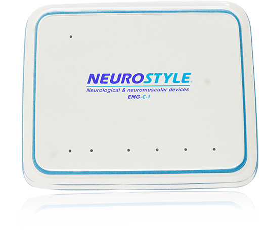
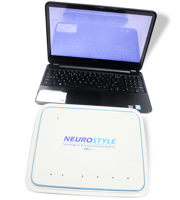
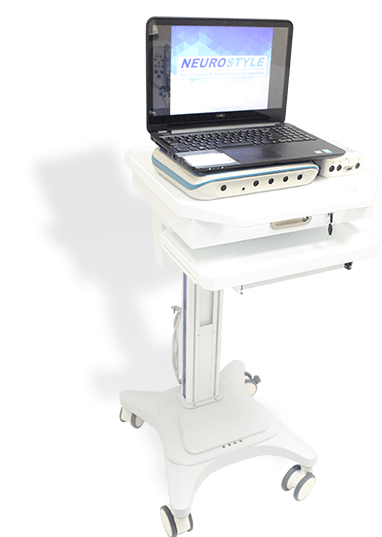
NS-EMG-C-1 is a digital EMG device. It acquires, stores and review up to 4 channels of EMG data and 2 channels evoked potential (EP) channels. It can perform routine EMG system recording, EP, as well as nerve conductive velocity (NCV) diagnostic examination.
EMG/EP
EMG System | EMG Device Key Features
- Pre-amplifier operable with up to 4 EMG channels
- High signal quality with USV data transmission, shielding the signal from power line interference
- Consists of 3 standard EMG modules with 4 EP modules
- Easy-to-understand illustations of electrode placements, stimulation positions and standard waveforms
- Easy access to waveform data comparison
- Ability to review and generate report for past cases


- Display Sensitivity: 0.05μV/div ~ 20,000μV/div, ±10% error. No precision requirements below 50μV/div.
- Frequency-Amplitude Characteristics: 0.2Hz ~10 kHz, error is +10% ~ -50% between 0.2Hz ~ ≤0.5Hz, and error is +5% ~ -30% between >0.5Hz ~10 kHz.
- Scan Speed: 1ms/div ~ 5000ms/div; error = ±10%;
- High-Pass Filter: 10Hz ~ 20,000Hz; error = +10% ~ -30%;
- Low-Pass Filter: 0.1Hz ~ 5000Hz; error = +10% ~ -30%.
- Common Mode Rejection Ratio: ≥110dB;
- Input Impedance: ≥1000MΩ;
- Noise Voltage:= 0.4μV (RMS);
- Resistance to Polarization Voltage: At ±300 mV DC polarization voltage, bias is ≤5%.
- Current Pulse Output Intensity: Maximum output 100mA; at 0mA ~ 4mA, step difference is 0.01mA. Error = ±10%;
- Pulse Output Frequency: 0.1Hz ~120Hz, error = ±10%;
- Target Pulse Width: 50μS, 100μS, 200μS, 300μS, 500μS, 1000μS; error = ±10%;
- Stimulus Direction: Positive, Negative, and Two-Way.
A. Normal Mode (Acoustic Stimulation) Parameters
- Stimulation Type: Short Tone, Pure Tone, White Noise.
- Stimulation Frequency: 0.1Hz ~100Hz, error = ±10%;
- Target Signal Probability: 5% ~ 100%, error = ±10%;
- Stimulation Model (for Short Tone and Pure Tone only): Left Ear, Right Ear, Both Ears, Alternate Ears.
- Short Tone Only
- Short Tone Stimulation Phase: Sparse Wave, Dense Wave, Alternating Waves;
- Maximum Short Tone Intensity: approx. 130dB;
- Pure Tone only
- Maximum Pure Sound Intensity: approx. 120dB;
- Pure Tone Sound Frequency: 250Hz ~7kHz, error = ±10%;
- White Noise only
- Maximum White Noise Intensity: approx. 100dB;
B. Multi-Frequency Auditory Steady-State Response (ASSR) Audio Stimulation Parameters
- Modulation Type: AM/FM
- Audio Stimulation Intensity: -10dB ~ 130dB HL (Hearing Level), error within ±10%;
- Carrier Frequency: 250Hz ~ 8000 Hz, error within ±10%;
- Modulation Frequency: 20 ~ 200 Hz, error within ±20%;
- Degree of AM: 0 ~ 100%, error within ±30%;
- Degree of FM: 0 ~ 30%, error within ±30%;
- AM/FM Phase Difference: 0 ~ 359 degrees, error within ±30%;
C. Video Control Output Signal (Visual Stimulation)
- Checkerboard Image
- Monitor can be set to display full-screen checkerboard image with alternating black-and-white tiles.
- Checkerboard display can be 4X3, 8X6, 16X12, 32X24, or 64X48.
- Half-screen checkerboard display:
- Left-half or right-half of screen: 2X3, 4X6, 8X12, 16X24, or 32X48 pattern,
- Upper or lower half of screen: 4X2, 8X3, 16X6, 32X12, or 64X24 pattern
- Quarter checkerboard display: 2X2, 4X3, 8X6, 16X12, or 32X24 checkerboard pattern.
- Horizontal Bar Grid Image
- Full-screen black-and-white horizontal bar image: 1X3, 1X6, 1X12, 1X24 and 1X48
- Left-half or right-half of screen only: 1X3, 1X6, 1X12, 1X24 and 1X48;
- Top or bottom half of screen only: 1X2, 1X3, 1X6, 1X12 and 1X24,
- Quarter screen display: 1X2, 1X3, 1X6, 1X12 and 1X24.
- Vertical Bar Grid Image
- Full-screen black-and-white alternating vertical bars: 4X1, 8X1, 16X1, 32X1, and 64X1
- Left half or right-half of screen only: 2X1, 4X1, 8X1, 16X1 and 32X1
- Top or bottom half of screen only: 4X1, 8X1, 16X1, 32X1 and 64X1
- Quarter screen display: 2X1, 4X1, 8X1, 16X1, and 32X1.
- Stimulation Frequency: 0.1Hz – 1Hz, error = ± 10 %;
- Target Signal Probability (P300 only): 5% ~ 100%, error = ± 10 %;
- Non-target signal 1 Probability (P300 only): 5% ~ 100%, error = ± 10 %;
- Non-target signal 2 Probability (P300 only): 5% ~ 100%, error = ± 10 %.
D. Goggles (Visual Stimulation)
- Stimulus Frequency: 0.1Hz – 50Hz, error = ± 10 %;
- Stimulation Modes: Left Eye, Right Eye, Both Eyes, Alternate Eyes.
System Key Features:
- Pre-amplifier operable with up to 4 EMG channels
- High signal quality with USB data transmission, shielding the signal from power line interference
- Consists of 3 standard EMG modules and 8 NCV modules
- Easy-to-understand illustrations of electrode placements, stimulation positions and standard waveforms
- Easy access to waveform data comparison
- Ability to review and generate report for past cases
Nerve Conduction Velocity (NCV)
- Waveform caching function enables saving of multiple waveforms with the same examination point
- Screen display of individual F-wave, H-wave and overlay
- Display of latency and amplitude values with measurement lines for easy reference


System Parameters:
A. Amplifier Parameters
- Display Sensitivity: 0.05μV/div ~ 20,000μV/div, ±10% error. No precision requirements below 50μV/div.
- Frequency-Amplitude Characteristics: 0.2Hz ~10 kHz, error is +10% ~ -50% between 0.2Hz ~ ≤0.5Hz, and error is +5% ~ -30% between >0.5Hz ~10 kHz.
- Scan Speed: 1ms/div ~ 5000ms/div; error = ±10%;
- High-Pass Filter: 10Hz ~ 20,000Hz; error = +10% ~ -30%;
- Low-Pass Filter: 0.1Hz ~ 5000Hz; error = +10% ~ -30%.
- Common Mode Rejection Ratio: ≥110dB;
- Input Impedance: ≥1000MΩ;
- Noise Voltage:= 0.4μV (RMS);
- Resistance to Polarization Voltage: At ±300 mV DC polarization voltage, bias is ≤5%.
B. Current Stimulation Parameters
- Current Pulse Output Intensity: Maximum output 100mA; at 0mA ~ 4mA, step difference is 0.01mA. Error = ±10%;
- Pulse Output Frequency: 0.1Hz ~120Hz, error = ±10%;
- Target Pulse Width: 50μs, 100μs, 200μs, 300μs, 500μs, 1000μs; error = ±10%;
- Stimulus Direction: Positive, Negative, and Two-Way.
EMG machine uses
- Electromyography (EMG) machines are largely used in clinical settings or hospitals to monitor muscles cells while they are active and at rest.
- The EMG machines are widely used as a research tool for studying kinesiology, motor control disorders.
- The EMG signals can be used to activate or regulate external assistive devices such as orthoses and prosthesis.
- With the advancement of technology the EMG technique and signals are used for neuropsychological mapping during brain surgery, it helps in monitoring the eloquent areas.
- Electromyography is being used in a myriad of evolutionary ways like in engineering applications. The myoelectric signal produces activation magnitude, that magnitude is then used to deduce a target force and motion of a worn mechanical appliance emulsion.
Find a range of high quality medical devices such as “eeg system” & “iom system” at Neurostyle
