Professional Intra-operative Neural-physiological Monitoring (PIOM) System

Model: PIOM-XP

By monitoring evoked potentials, EMG, TOF and other items, it provides surgeons with objective evaluation indicators of central nerves, peripheral nerves, muscles and narcotics, provides real-time feedback on the activity of nerves during surgery, and guides surgeons (e.g. neurosurgery, orthopedics, ENT, vascular surgery, thyroid surgery, thoracic surgery, etc.) on whether nerves are touched during the progress of surgery or whether there is any damage to nerves and the site of the damage.
PIOM SYSTEM TECHNICAL SPECIFICATIONS
AMPLIFIER
• Number of channels: amplifier 16/ 32 channels.
• Supports up to 32 channels of signal acquisition, with multiple channels integrated into a single output port for connection to consumables, and supports quick connection of acquisition electrodes.
• Noise level: :::; 2.0 µV p-p (0.2-200Hz)
• Input impedance: 3000M
• With filtering function: high frequency filtering, 1 0HZ-7000HZ, low frequency filtering 0.1-500Hz, multi-position adjustable.
• Common mode rejection ratio 120dB.prevent accidental vocal cord injury, and the electrode placement ring is designed for improved muscle contact.
ELECTRICAL STIMULATOR
- Stimulus polarity: positive phase, negative phase, biphasic
- Stimulation pulse width: 0.025 – 1.0 ms
- System current stimulation intensity: maximum stimulation voltage 1000 V, maximum output current 1000 mA
- Pulse string frequency: 1Hz-1000Hz.
- Constant current stimulator: .:: 9 high current stimulation ports
ACOUSTIC STIMULATOR
- Stimulus type: Short tone / pure tone /white noise
- Stimulation output: left ear, right ear, both ears simultaneously, alternating between ears.
- Maximum short tone/ pure tone/ white noise Sound intensity :5 125dB
- Stimulation frequency: 0.1 Hz-1 00Hz
PIOM SYSTEM SOFTWARE FEATURES
- Computer operating system software: Windows 10 Pro system, professional version of Office software.
- Nerve monitoring software items: EEG, EMG, cortical localization, somatosensory evoked po-tentials(SEP), motor evoked potentials(MEP), automatic vertebral pedicle stimulation program, near nerve detection function, auditory evoked potentials(AEP), visual evoked potentials (VEP), etc.
- Multiple inspections are carried out simultaneously on the same screen and can be switched freely.
- Various monitoring modes for all modules can be edited and added according to the needs of the operation, and can be saved as a template to facilitate future monitoring of similar opera-tions.
- TOF test function.
- Noise analysis shielding software.
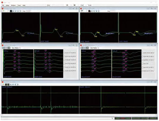
- With multiple data windows display: Real-time waveforms, trend graphs, data tables, video images, event windows, etc., it can be displayed on the same screen, or can be viewed window by window in separate screens.
- Report: Template function, can generate report with one click, user can edit and save it by themselves.
- Screen printing function: The monitoring waveform can be copied to the screen and automati-cally imported into the report or saved as a picture format.
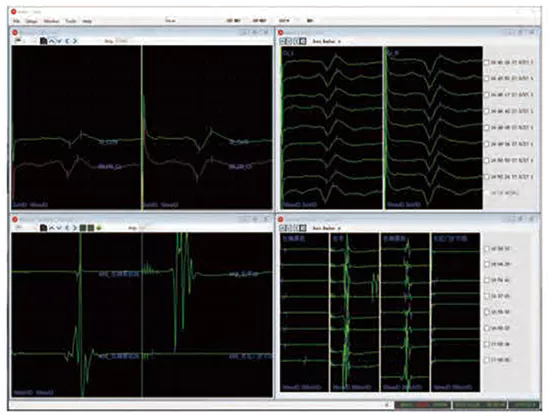
Nerve Signal Monitoring
Real-time monitoring with mode and stimulation controls ..

DISPOSABLE NEEDLE ELECTRODE
Integrated design of the consumables
Multiple consumables are integrated into one connector and can be connected in one go.
Help reduce workload and almost achieve zero -error operation during connection process.
ANTI-INTERFERENCE CAPABILITY
Anti-interference cable of integrated
Consumables greatly increase the resistance of interference, avoiding interference with origi- nal waveform, increasing the fidelity of the waveform,avoiding misjudgment caused by excessive interference.
SPIRAL ELECTRODE
Screw into patient’s scalp, are firmly attached and not easily dislodged.
lntraoperative stimulation current output, acting on motor evoked potential monitoring.


SURFACE ELECTRODE
Patch type surface electrode, apply to the skin surface of near nerves, e.g. median nerve, tibial nerve.
lntraoperative stimulation current output, acting on somatosensory evoked potential monitoring.
DISPOSABLE NERVE PROBE
Dedicated surgeon hand-held probe, with current output at the tip. It can be used to stimulate uncertain body tissue during surgery to assist in judging nerve alignment, or directly acting on pedicle screw, adjusting nail placement angle according to the feedback of current parameters.

Technical Parameters of the Intra-operative Nerve Monitor System
Main Functions: by monitoring evoked potentials, EMG, TOF and other items, it provides surgeons with objective evaluation indicators of central nerves, peripheral nerves, muscles and narcotics, provides real-time feedback on the activity of nerves during surgery, and guides surgeons (e.g. neurosurgery, orthopedics, ENT, vascular surgery, thyroid surgery, thoracic surgery, etc.) on whether nerves are touched during the progress of surgery or whether there is any damage to nerves and the site of the damage.
Technical Specifications: main technical specifications, parameters.
Amplifier
- Number of channels: amplifier 16/ 32 channels.
*2. Supports up to 32 channels of signal acquisition, with multiple channels integrated into a single output port for connection to consumables, and supports quick connection of acquisition electrodes.
- A/D conversion ≥ 24 bit
- Noise level: ≤ 2.0 μV p-p (0.2-200Hz)
- Sensitivity: 0.1μV/D to 30mV/div step control
*6. Input impedance: ≥ 3000MW
- Impedance measurement: all input electrodes can be detected real-time
- With filtering function: high frequency filtering, 10HZ-7000HZ, low frequency filtering 0.1-500Hz, multi-position adjustable
- Common mode rejection ratio ≥ 120dB
- Isolation power supply: equipped with special medical isolation power supply, anti-hyperbaric special isolation.
Electrical Stimulator
- Constant current and voltage, it can’t be achieved with the external third-party stimulator.
- Output modes: repetitive, non-repetitive, single pulse train
- Safety: power limit, power on test
- Stimulus polarity: positive phase, negative phase, biphasic
- Stimulation pulse width: 0.025 – 1.0 ms
*6. System current stimulation intensity: maximum stimulation voltage 1000 V, maximum output current 1000 mA
- Pulse string frequency: 1Hz-1000Hz
- Constant current stimulator: ≥ 9 high current stimulation ports
Acoustic Stimulator
- Stimulus type: short tone / pure tone / white noise
- Stimulation output: left ear, right ear, both ears simultaneously, alternating between ears.
- Maximum short tone sound intensity ≤ 125dB
- Maximum pure tone sound intensity ≤125dB
- Maximum white noise sound intensity ≤125dB
- Stimulation frequency: 1Hz-100Hz
Visual Stimulator
- Checkerboard grid image
The display can show a full screen black and white flipped checkerboard grid image with the following numbers: 4 x 3, 8 x 6, 16 x 12, 32 x 24, 64 x 48. It can also show 2 x 3,4 x 6, 8 x 12, 16 x 24, 32 x 48 checkerboard grids on the left half screen and right half screen respectively; 4 x 2, 8 x 3, 16 x 6, 32 x 12, 64 x 24 on the upper half screen and lower half screen separately; In the first quadrant, second quadrant, third quadrant, fourth quadrant displaying 2 x 2, 4 x 3, 8 x 6, 16 x 12, 32 x 24 checkerboard grids.
- Horizontal bar grid image
The display can show a full screen black and white flipped horizontal bar image, the number of bars can be set to 3, 6, 12, 24, 48. It can also display 3, 6, 12, 24, 48 bars in the left half of the screen and the right half of the screen respectively; In the upper half of the screen and the lower half of the screen display 3, 6, 12, 24 horizontal bar grid respectively; In the first quadrant, the second quadrant, the third quadrant, the fourth quadrant display 2, 3, 6, 12, 24 horizontal bars grid.
- Vertical bar image
The monitor can display a full screen black and white flipped vertical bar image, the number of display is 4, 8, 16, 32, 64 separately. It can also display 2, 4, 8, 16, 32 vertical bars in the left half of the screen, right half of the screen; In the upper half of the screen, the lower half of the screen display 4, 8, 16, 32, 64 vertical bars; In the first quadrant, the second quadrant, the third quadrant, the fourth quadrant display 2, 4, 8, 16, 32 vertical bars.
- Stimulation frequency: 0.1 Hz to 1 Hz, error not exceeding ±10%.
- Target signal probability: 5% to 100%, target probability error not more than ±10%.
- Flash stimulation
Stimulation frequency: 0.1Hz~50Hz, error not more than ±10%.
Stimulation mode: left eye stimulation, right eye stimulation, simultaneous stimulation of both eyes, alternate stimulation.
Software Features
- Computer operating system software: Windows 10 Pro system, professional version of Office software.
*2. Nerve monitoring software items: EEG, EMG, cortical localization, somatosensory evoked potentials(SEP), motor evoked potentials(MEP), automatic vertebral pedicle stimulation program, near nerve detection function, auditory evoked potentials(AEP), visual evoked potentials(VEP), etc.
- Multiple items can be monitored simultaneously, simultaneous parallel monitoring of EEG, evoked potentials and EMG, all-round monitoring of functional nerves at risk during surgery.
- Multiple inspections are carried out simultaneously on the same screen and can be switched freely; the inspection order function can be set freely, and different monitoring order inspections can be carried out at certain intervals.
- TOF test function: the value of the attenuation of each waveform can be measured directly and fully automatically, and displayed in a histogram for more intuitive display.
* 6. EEG functions: Spectrogram: The spectrum analysis of waveform data calculates the relative energy share of each frequency band δ, θ, α, β, the middle frequency index, the side frequency index, the relative energy ratio of fast and slow waves, the burst inhibition ratio, the amplitude integration, and the dual frequency index for the whole case, which is expressed in the form of a trend graph.
- EMG function: consists of free EMG waveform window, trigger EMG window and EMG waveform stack window, which displays the collected EMG waveform data in real time and can be temporarily played back. Switchable single-area and horizontal tiled dual-area display window layout types. Automatic capture of EMG action unit potentials, including single stimulus-triggered data capture. Save with waveform Label, capture time, capture source etc.
- Evoked potential function: three ways to display waveform data in real time: (1). absolute value of data;
- difference between each waveform and baseline;
- percentage data between each waveform and
- Noise analysis shielding software: software with noise analysis function, can analyze the operating room bipolar electrocoagulation and other noise, shielding acquisition.
- With multiple data windows display: real-time waveforms, trend graphs, data tables, video images, event windows, , it can be displayed on the same screen, or can be viewed window by window in separate screens.
- Monitoring template editing and saving: with various monitoring modes, you can edit and add monitoring modes according to different surgical methods with unlimited numbers. The system provides several templates of monitoring programme parameters that have been configured. The user can select a certain template parameter to become the parameter of the current monitoring programme, and the user’s modified monitoring programme parameters can also be saved as a new
- Real-time monitoring software: displays the patient’s muscle relaxation; calibrates the signal quality and displays disturbances in real time, facilitating timely adjustment of the operation.
- TOF monitoring: shows the patient’s muscle relaxation
- Report: template function, can generate report with one click, user can edit and save it by themselves, support English report, compatible with word document processing software, each display windows can be copied and pasted to other applications.
- Screen printing function: the monitoring waveform can be copied to the screen and automatically imported into the report or saved as a picture format.
- Any computer can be used for instant network functions via LAN or VPN.
- The device has an automatic storage function and can be picked up after an accidental power failure for continuous monitoring without the need to start again.
- Automatic pedicle of vertebra test function with automatic adjustment.
- With a near nerve detection function, it is convenient to know the distance between the stimulation point and the nerve.