The electromyography or EMG is an electrodiagnostic test that is performed to test and assess the health of skeletal muscles and motor neurons — the nerve cells that control the muscles.
The motor neurons send electrical signals through them that cause the muscles to contract and relax. Electromyography or EMG system measures this electrical activity in muscle in response to nerve’s stimulation of the muscle and convert these signals into graphs or numerical values, helping specialists or doctors interpret them to make a diagnosis
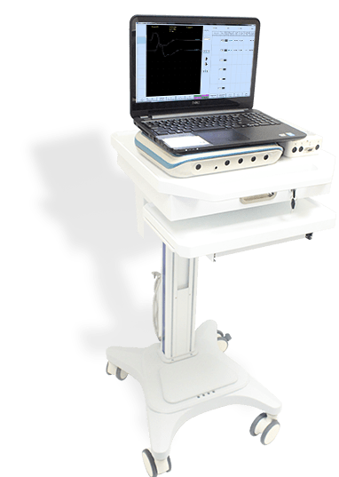
EMG systems produced nowadays are computer based which includes dedicated hardware units like a stimulator, amplifier, preamplifier, control panel, speaker, computer, printer, and several types of electrodes. The modular configuration of modern EMG systems makes it possible to upgrade or replace the computer, while keeping the other hardware units intact. The most important part of an EMG system is its amplifiers and stimulators.

Why would someone need an EMG? / Why is electromyography performed?
Usually when a doctor sees symptoms and suspects muscle or nerve disorder — he will order an EMG. Few symptoms may include numbness, tingling, muscle pain or cramping, unexplained weakness in the limbs or certain types of pain in limbs. EMG scan results are then used to detect muscle dysfunction, nerve dysfunction, or problems with signal transmission from nerve-to-muscle. The possible causes when EMG is required may include:
- Muscle weakness due to disorders like muscular dystrophy or polymyositis
- Skeletal muscle weakness because of a problem in motor neuron signal transmission to the muscle such as myasthenia gravis
- Numbness, weakness or unexplained pain which may be for ‘pinched nerve’ in the spine or radiculopathy
- Numbness, tingling or pain in thumb or first three fingers of hand, which may be because of peripheral nerve disorders that affect nerves outside spinal cord such as peripheral neuropathies or carpal tunnel syndrome.
- Degenerative motor nerve disorders like amyotrophic lateral sclerosis (ALS) or polio
- Disorders that affect root of the nerve like herniated disk in the spine
Apart from the above causes, there are several other reasons for which EMG test may be required.
Is EMG test painful? / Why was my EMG so painful? / How painful is an EMG test?
Generally, patients have different levels of understanding and perceptions of EMG. But only few are well-informed. It will be helpful giving information before the test.
Few people ask “Is EMG test painful?” Some level of discomfort may be felt by the patient when the electrode or stimulator needle is inserted. The needles will be attached to a EEG machine by wires. But it is usually painless and well tolerated, with no pain medication. If the test is painful, the examiner must be notified immediately because this can interfere with the results. The pain during EMG may be eased with pharmacologic interventions such as skin sprays or oral analgesics, and with non-pharmacologic interventions such as calming music or providing pre-procedural information about the test to ease anxiety.
EMG testing has two parts, and both may cause some discomfort. But they are usually well tolerated, which ends shortly after the needle is removed. In most cases, the doctor will perform both parts and in some situations anyone may be done. The two parts of the EMG test are:
- Nerve conduction study: Specific nerves are stimulated at different points with small electric shocks, and their function — ability to send the impulse to the muscle is recorded and measured.
- Needle examination for muscle testing — Very fine needles containing microscopic electrodes are inserted into several muscles. Each needle picks up both the normal and abnormal electric signals emitted by those specific muscles when contracted and in rest.
How do you prepare for an EMG? / How long does an EMG test take?
No special preparation is required. Restrain from smoking for at least three hours before the test will be conducted. Fasting is not required during the test. Don’t apply any creams or lotion before EEG test. But the below points must be followed in some cases:
- Smoking, caffeinated beverages or certain drugs that act on the nervous system (such as muscle relaxants) can interfere with EMG results will be restricted three hours to six days before the test.
- Tell the EMG technician or doctor about all prescribed or over-the-counter and herbal supplements which the patient may take.
- Notify the doctor if the patient has bleeding disorder and taking blood-thinning drugs, or if the patient has an implantable defibrillator or a pacemaker.
- Don’t apply any creams or lotion before EEG test.
- Based on one’s medical condition, the doctor may advise other specific preparation.
What you can expect? / What happens during an electromyography EMG?
The first part of the procedure is Nerve Conduction Study (NCS). During the study, small sensors called surface electrodes are placed on the skin to assess the ability of the motor neurons to send electrical signals.
Before the Nerve Conduction Study (NCS) procedure:
- The patient will be asked to remove jewelry, hairpins, hearing aids, eyeglasses, or other metal object that can interfere with EMG procedure. Also, likely to be asked to remove cloths and change into a hospital gown and sit on a reclined chair or lie down on an examination table for the test procedure.
- A neurologist or technician will the specific nerves for the test to conduct and place recording and stimulating electrodes over the nerve on the skin with special pastes. Sometimes the neurologist may electrode needles at different spots, depending on the symptoms.
During the NCS procedure:
- The surface stimulating electrodes will transmit mild and brief electric current to stimulate the nerve. Some minor discomfort can be experienced for a few seconds as this may feel as a spasm or twinge.
- The signals or responses from the motor nerves to-muscles after stimulation will be detected by the electrodes, then recorded and displayed on a monitor for further assessment by the neurologist.
After the NCS procedure:
- On completion of the test, the electrodes will be removed from the skin and the paste used to attach will be washed off.
- After the test, there may be some temporary minor bruising at some spots which shall fade away within a few days. Unless the doctor advises differently after the procedure, which depends on individual situation, the patient may return to normal activities
Needle examination or needle EMG is the second part of the EMG procedure. This process also uses sensors called needle neurons to test electrical signals. The sensors are directly inserted into tissue of affected muscle to evaluate muscle activity when at rest and when contracted.
During the Needle EMG procedure:
After the Nerve Conduction Study (NCS), the neurologist or doctor will perform the needle EMG where he will assess the natural electrical activity when the muscle is at both rest and contracted position.
- The skin on the affected area will be first cleaned with an antiseptic solution by the doctor. After that, using a fine sterile needle, electrodes will be inserted into the muscle tissue. And a ground electrode is placed under the arm or leg.
- Some times five or more electrodes are required to be inserted for the needle examination procedure and a slight bearable discomfort may be felt while the needle is being inserted. The pain from needle insertion will subside shortly once the needles are removed after the test is over.
- The doctor will give instructions to relax and then slightly or with full strength to contract muscles at an appropriate time. According to which muscles and nerves are examined, he may ask to change positions during exams.
- If the discomfort becomes unbearable or painful, the doctor must be notified because that may interfere with the test results.
- Then the electrical activity from the working muscles, when contracted and when at rest, will be detected by the electrodes, then recorded and displayed on a monitor for further assessment by the doctor.
After Needle examination procedure:
- Once the test is over the electrodes will be removed.
- The patient may experience some temporary muscle soreness or minor bruising where the needle will be inserted for a day or so following the procedure.
- The soreness or bruising should fade away within several days. If it persists, pain increases and it swells or pus you may notify your doctor or primary healthcare.
Results / What happens if EMG is abnormal?
The neurologist or doctor will review and interpret the results right after the procedure and will prepare a report. Although if any other doctor ordered the EMG scan, the result may not be known until a follow-up appointment is arranged with the doctor.
Depending on the results, the doctor will discuss the report and about additional treatments or tests that might be required at a follow-up appointment.